> Research & Science
NanoSIMS
Imaging techniques gain popularity in analytical and biological sciences. Already in the 16th and 17th centuries, light microscopes gave access to biological cells. Nowadays, powerful electron microscopes, notably transmission electron microscopes (TEM), allow the imaging at micro and nanometer levels, from subcellular structure down the atoms.
There is growing interest in coupling bio-imaging with chemical information. TEM can be combined with energy-dispersive X-ray spectroscopy (TEM/X-EDS), but its element sensitivity is only in the percentage range. Laser Ablation (LA) ICP-MS generate elemental images from biological tissues, but its spatial resolution is limited at the micrometer level, which is not sufficient for subcellular imaging.
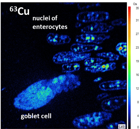
Secondary ion mass spectrometry (SIMS) has been increasingly recognized as a powerful technique for visualizing molecular architectures in cell biology. SIMS provides high spatial resolution down to 50-100 nm, which is sufficient for the observation of subcellular structures, combined with element sensitivity at the mg/kg level. From all the SIMS techniques, nanoSIMS provides unsurpassed lateral resolution.


Although the number of NanoSIMS instruments worldwide that are dedicated to biological applications is relatively limited, it offers a wide range of applications for research on trace minerals in animal tissues.
Source: Schaumlöffel, D. (2020), Secondary Ion Mass Spectrometry for Biological Applications, J. Anal. At. Spectrom., 35, 1045-1046
